Contributing author: Iris Friedli, Senior Director MR Imaging @ Antaros Medical
The Magnetic Resonance Imaging (MRI) field is constantly evolving, particularly with respect to what is possible in the research setting.
It is always exciting to attend the International Society for Magnetic Resonance in Medicine (ISMRM) annual meeting and exhibition and get a snapshot into where the MRI field is heading. This year’s meeting in Toronto was no exception, and I felt as though the programme and the surrounding discussions reflected a shift in the way we are thinking about MRI, its value as an imaging technique in different settings, and the current challenges being faced by its users globally.
As my work with MRI is in the context of clinical trials and drug development, it is easy for me to forget that this is not always the biggest focus in the field of MRI. However, a notable addition at this year’s meeting was the session “Clinical Trials Demystified: Who are all the stakeholders when MRI is used?”, which focused on how MRI can be used in clinical trials. It was great to see a growing interest in the value MRI can bring to drug development.
While MRI principles are largely unchanged, technological advances in both the hardware and software of scanners, as well as changing societal needs, are continually furthering the possibilities for MRI as an imaging technique. In this blog post I want to briefly discuss some of the latest innovations to hardware (advances in magnetic fields and sustainability), software (integration of artificial intelligence), and some of the societal needs that were highlighted during ISMRM (ethics and global availability) and will have important implications for the use of MRI in clinical trials and drug development.
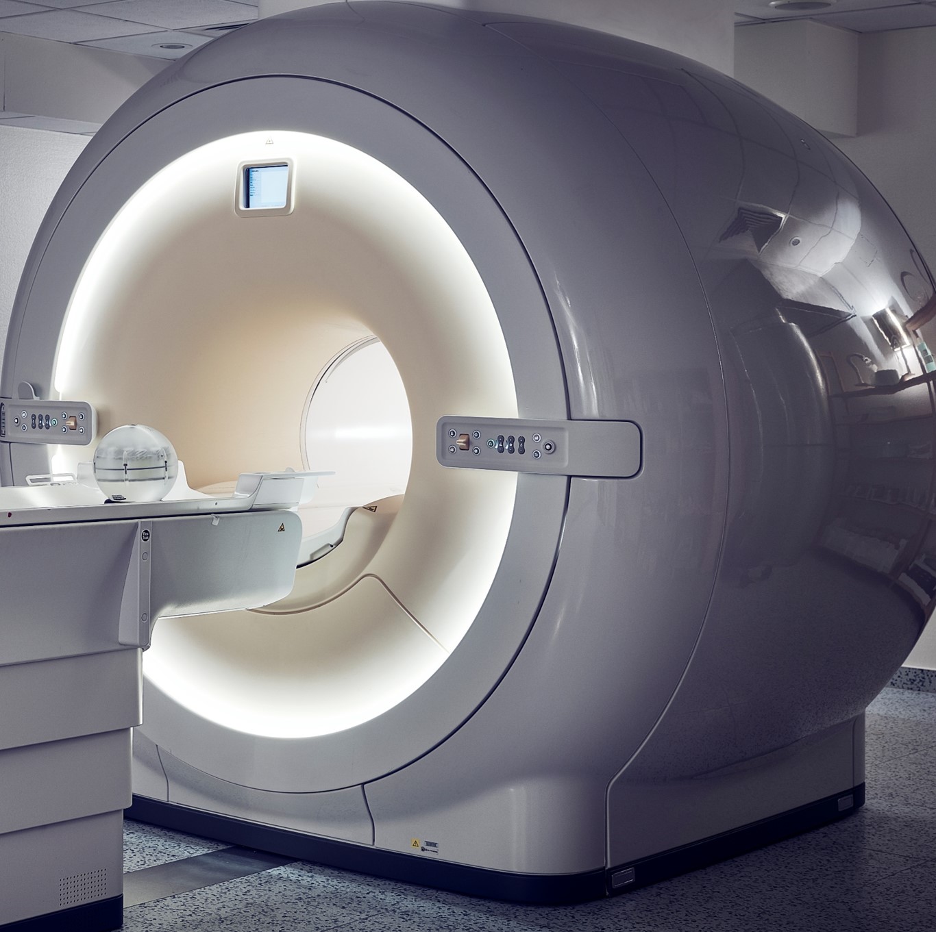
What is MRI and what’s advancing in MRI hardware?
MRI, in short, uses a strong magnetic field to align protons in the same direction and then sends out a radio wave, or pulse, which causes the protons to move. The radiofrequency (RF) coils transmit the pulse, producing a small magnetic field perpendicular to the main magnetic field which moves the protons. When the radio pulse is removed, the protons revert to their original position and the signal that is created from this is detected by the receiver coils and reconstructed into an image by a computer. A ‘scan’ usually involves a sequence of radio pulses, the timing and duration of which can vary depending on the question of interest.

Higher magnetic fields for more detailed images
Scanners are generally referred to with regards to the strength of the magnetic field in terms of ‘T’ or tesla, a unit of measurement used to describe magnetic flux density. The strength of the magnet directly affects the strength of the signal that can be collected from the scan and therefore the image quality.
The standard for most MRI scanners in a clinical setting is 1.5T or 3T which is suitable for most routine scans. However, there has long been a desire to have even stronger magnetic fields, particularly in research. For example, both 5T and 7T models were being showcased at ISMRM (by United Imaging and Siemens, respectively). The stronger the magnetic field, the stronger the signal, and the more detailed the images can be. This can be very advantageous in research, especially when there are questions regarding mechanism of action. To date, the strongest magnetic field I have heard of is a 14T scanner that is being developed by a research consortium in the Netherlands.
Open MRI scanners to accommodate more patients
Conversely, there have also been advances in open bore scanners with lower magnetic fields. MRI scanners are shaped like a tube, and the patient lies on a table in the opening of the tube, which is also called the bore. Sometimes referred to as ‘closed bore’, a standard scanner is closed on 3 sides and typically has a bore that is 60cm in diameter or 70 cm (‘wide bore’). Other scanners have been developed which sandwich the patient rather than closing them in (‘open MRI’).
The advantage of improved patient comfort in these types of scanners does come at the expense of requiring stronger magnetic fields. Most open MRI scanners have a magnetic field strength between 0.3T and 0.7T, however, at ISMRM, Fujifilm presented their 1.2T open bore scanner, which can be used to accommodate larger patients or those who suffer from claustrophobia. In the context of clinical trials and drug development, this could positively impact patient recruitment and enable inclusion of patients who would otherwise not be able to participate in a study.
Sustainability
The medical imaging field has recently begun to consider the environmental impact of imaging in the same way it has previously considered the potential dangers of radiation. There is room for improvement in many aspects. Two that are currently being focused on are reducing energy consumption and limiting the depletion of resources.
Maintaining the magnetic field is very energy intensive and generates a lot of heat. Within the MRI scanner, the magnet coil is housed in a bath of liquid helium to keep the temperature down, and energy is almost constantly required for this cooling. It has been estimated that over a one/year period, an MRI scanner will use as much energy as 26 four-person households, and approximately 60-70% of an MRI’s total energy consumption is used for the refrigeration of the magnet.
To reduce energy consumption, some manufacturers have developed built/in features such as sleep mode (also sometimes called economic power mode or EPM), during which the refrigeration is turned off and on in intervals, rather than constantly running.
Other sustainability concerns regarding MRI have centred around the use of helium in scanners. As the helium evaporates over time with repeated use, MRI scanners have historically required periodic helium refills. However, helium is a rate resource, and particularly amid recent helium shortages, the importance of finding solutions to conserve helium is well recognised. Newer scanners, such as those presented by GE HealthCare at ISMRM, use significantly less helium and recaptures used helium so that it doesn’t evaporate over time and will not need to be refilled.
How is artificial intelligence (AI) impacting the field of MRI?
There is no denying that artificial intelligence (AI) is impacting many different industries and technologies. Even within the field of MRI, it is being used in a variety of ways, however, today I will only discuss how it is being integrated to speed up the scanning process.
Acquiring an MR imaging from the detected signal involves several steps that each will have an impact on the quality of the image. Some of these steps are common across most reconstruction processes, but others can be customised for certain applications. One way to shorten the time required for a scan, without compromising on resolution, is to use an AI-based acceleration technique, such as those presented by Phillips and GE HealthCare at ISMRM. This can speed up the image acquisition and reconstruction process and significantly shorten scan times. Shorter scan times can have implications for patient recruitment, but also reduce the likelihood of patient non-compliance that might compromise the images.
Changing societal wants and needs of MRI
Two of largest challenges for MRI that were discussed at the meeting were the increasing concerns around ethics and global availability issues.
Ethics and data sharing
There was a large focus on ethics in MRI, more specifically around issues of sharing data, the need for open-source data, and issues around data quality. in the context of drug development, this is a particularly complex issue, as shared interests in drug development often don’t align with regulatory or privacy and security requirements.
Global availability issues
While in the United States and most of Europe the primary concern for improving MRI availability is reducing scan time and thereby the costs of seeing patients, there are very different issues facing the availability of MRI in different parts of the world.
For example, in Southeast Asia the main limitation to accessibility is the number of scanners, but there are also considerations regarding infrastructure demands such as helium availability or the ongoing operating costs of an MRI scanner. Whereas in Sub-Saharan Africa, it is the shortage of skilled personnel trained in MRI that is the biggest limiting factor. The Consortium for Advancement of MRI Education and Research in Africa (CAMERA) initiative is an example of the global efforts that are being made to address this by establishing a Pan-African interdisciplinary collaborative network of researchers and clinicals from 15 African countries.
In addition to its importance for patient care, scanner availability can also impact clinical trials. Plenty of work has been done to show the importance of geographical representation and cohort diversity in research, which highlights the importance of addressing these availability issues.
My main takeaway from ISMRM this year is that the field of MRI is constantly evolving, but also that it needs to in order to meet the changing requirements and needs of patients, healthcare providers, and researchers. I’m excited to see what is being presented next year and what progress is made between now and then.
In summary
To summarise briefly everything that has been discussed:
- Attending this year’s ISMRM gave me a snapshot into where the field of MRI is today and some of the challenges it is currently facing.
- Several technological hardware advances, such as stronger magnetic fields, open-bore MRIs and sustainable features are propelling MRI forward, especially in research.
- One of the ways that the integration of AI into MRI software is expanding the use of MRI is through shortening scanning times and speeding up reconstruction.
- Current societal needs such as ethical issues and global availability are also significantly influencing the way MRI is being used and developed.
Blog disclaimer
The views and opinions expressed in this article are solely those of the contributing author/s. These views and opinions do not necessarily represent those of Antaros Medical.
Contact details
If you have any questions regarding this article, please reach out to press@antarosmedical.com