Contributing author: Lars Johansson, Chief Scientific Officer at Antaros Medical
Understanding muscle composition is critical for interpreting treatment effects accurately. For example, loss of muscle fat should not be mistaken for loss of muscle volume, and specific muscle composition changes can have different effects on metabolic health, physical function and clinical outcomes.
Our understanding of adipose tissue within muscles, called intramuscular adipose tissue (IMAT, or intra-MAT) and its role in muscle health is evolving. A recent review paper by Jones et al. brings some long-needed clarity to IMAT definitions, what is currently known about its role in disease pathophysiology, imaging methodologies for the quantification of IMAT, and some directions for the future of this field.
This blog post will reflect on this publication and some of its implications for drug development and the quantification of IMAT as an endpoint in clinical trials.
Muscle fat compartments: clear definitions for a complex tissue
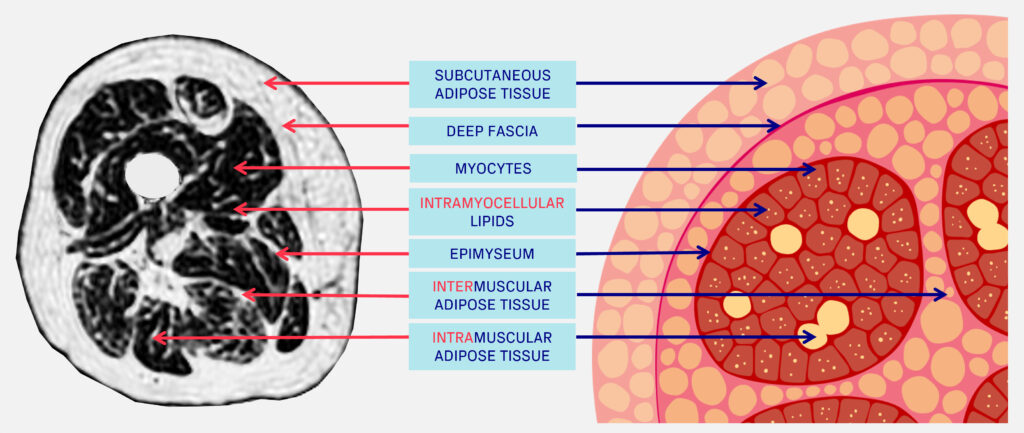
As Jones et al. note in this publication, there is a lack of consistency in definitions of adipose tissue related to muscles. They categorize muscle fat into the following:
- Intramuscular adipose tissue (intra-MAT): fat located within the muscle, expressed as volume or fat fraction. It includes fat located between muscle fibers and fascicles, within the epimysium.
- Intermuscular adipose tissue (inter-MAT): fat surrounding muscles beneath the deep fascial layer, outside the epimysium but between adjacent muscles.
- Intramyocellular lipids (IMCL): lipid droplets stored inside muscle cells, serving as an energy source but also associated with metabolic dysfunction when elevated.
The functional and metabolic consequences and contributions of IMAT
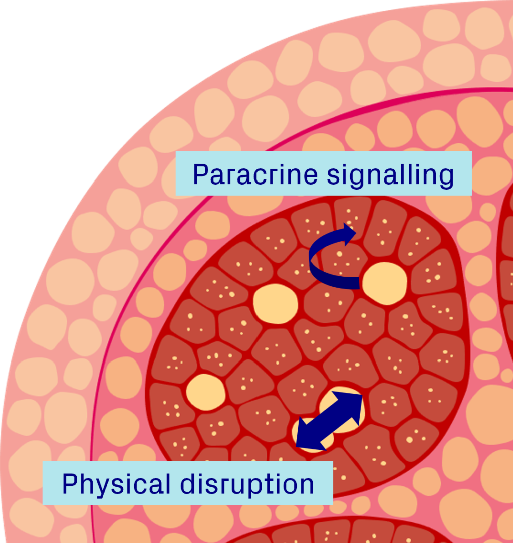
Decades of research has found associations between IMAT and a range of adverse clinical outcomes, impaired muscle function, and metabolic dysfunction. Exactly what underpins these links is slowly being revealed with emerging evidence. The authors propose that excess IMAT can negatively impact muscle function through both:
- Mechanical disruption: fat deposits within the muscle can physically separate individual muscle fibers, disrupting their coordinated activation, reducing muscle efficiency and overall contractile performance.
- Biochemical interference: IMAT releases adipokines that signal to nearby myocytes through paracrine pathways. These signals may promote inflammation, impair insulin sensitivity, and contribute to muscle degeneration.
Quantifying IMAT: Imaging methods and measurement approaches
The different muscle fat compartments are anatomically and functionally distinct and can be measured using imaging. Computed tomography (CT) or magnetic resonance imaging (MRI) are the most common ways of quantifying IMAT, however, to date this has been done inconsistently and in a variety of different ways.
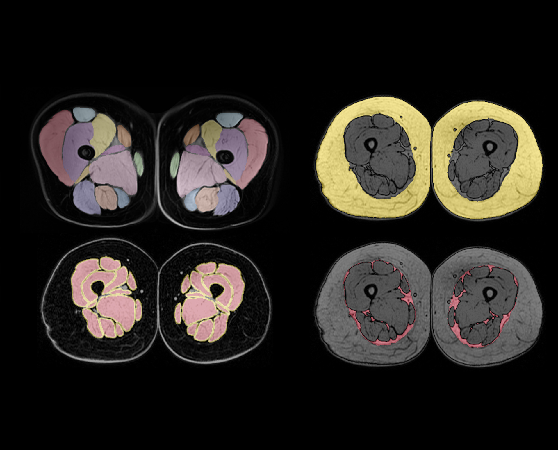
While it is also possible to use a thresholding approach, meaning that the differentiation between muscle and fat is determined by setting a threshold of signal intensity, at Antaros Medical we believe in segmenting all muscle and adipose tissue compartments. This is increasingly important in interventional trials where precise and accurate measurements are necessary to ensure that observed differences reflect true physiological changes rather than methodological variation, all of which is essential when interpreting treatment effects in clinical trials.
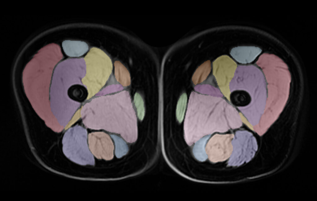
Imaging has also shown that the distribution and composition of IMAT is heterogeneous by muscle, disease, sex, and age. This becomes especially important when interpreting changes in muscle composition in relation to treatment interventions in clinical trials.
Moving the field forward: future directions for IMAT research
In addition to a call for standardization of definitions and increased transparency surrounding quantification methodologies, Jones et al. highlight another very interesting avenue for future IMAT research – seeking to answer the question: at what level of IMAT do we see functional deficit?
The authors, in previous preclinical work, showed that functional deficits became evident once IMAT levels reach 12% of the muscle area. This would be very interesting to look at in clinical trials and fits nicely with a growing body of research linking IMAT to different measures of muscle function.
Closing thoughts
This review by Jones et al. offers a timely and comprehensive overview of what is currently known about IMAT its biological relevance and its measurement complexities. The authors highlight the need for standardized definitions, greater transparency and consistency in quantification using imaging methods, and careful consideration of IMAT’s heterogeneity when interpreting longitudinal changes, particularly in interventional clinical trials. As the field increasingly recognizes the importance of IMAT in muscle health, these recommendations provide a valuable foundation for future research.
Blog disclaimer
The views and opinions expressed in this article are solely those of the contributing author/s. These views and opinions do not necessarily represent those of Antaros Medical.
Contact details
If you have any questions regarding this article, please reach out to press@antarosmedical.com